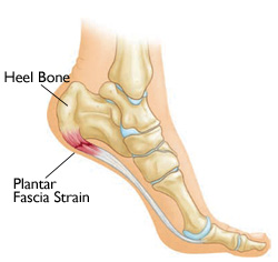
Plantar fasciitis is a painful inflammatory process of the plantar fascia. Longstanding cases of plantar fasciitis often demonstrate more degenerative changes than inflammatory changes and is termed, plantar fasciosis. The plantar fascia is a thick fibrous band of tissue originating on the bottom surface of the calcaneus (heel bone) and extending along the sole of the foot towards the five toes. The term fasciitis is a misnomer as studies of the tissue do not demonstrate inflammation. Plantar fasciosis is a more accurate diagnosis. It has been reported plantar fasciitis occurs in two million Americans a year and 10% of the population over a lifetime. It is commonly associated with long periods of work-related weight bearing. Among non-athletic populations, it is associated with a high body mass index. The pain is usually felt on the underside of the heel and is often most intense with the first steps of the day. Another symptom is that the sufferer has difficulty bending the foot so that the toes are brought toward the shin (decreased dorsiflexion of the ankle). A symptom commonly recognized among sufferers of plantar fasciitis is increased probability of knee pains, especially among runners.
Diagnosis
The diagnosis of plantar fasciitis is usually made by clinical examination alone. Imaging studies may include radiographs, diagnostic ultrasound and MRI. An incidental finding associated with this condition is a heel spur, a small bony calcification, on the calcaneus heel bone, in which case it is the underlying condition, and not the spur itself, which produces the pain.
Treatment
Non-surgical
While evidence is lacking to strongly support any type of treatment for plantar fasciitis, many non-surgical treatments are available. Stretching of the calf and plantar fascia can provide up to 2–4 months of benefit. In cases of chronic plantar fasciitis of at least 10 months’ duration, one study has shown high success rates with a stretch of the plantar fascia. Most often plantar fasciitis improves within one year of beginning non-surgical treatment, without any long-term problems.
Pain with the first steps of the day can be markedly reduced by stretching the plantar fascia and Achilles tendon before getting out of bed. Night splints can be used to keep the foot in a dorsi-flexed position during sleep to improve calf muscle flexibility and decrease pain on waking. These have many different designs. The type of splint has not been shown to affect outcomes. Arch supports and taping can provide short-term pain relief. There has not been shown to be any difference between custom-made orthotics and off-the-shelf orthotics in the treatment in acute plantar fasciitis despite the long term efficacy of custom made orthotics in chronic plantar fasciitis.
To relieve pain and inflammation, nonsteroidal anti-inflammatory drugs (NSAIDs) such as aspirin and ibuprofen are often used but are of limited benefit. Dexamethasone 0.4 % or acetic acid 5% delivered by ionophoresis combined with low dye strapping and calf stretching has been shown to provide short term pain relief and increased function.
Local injection of corticosteroids often gives temporary or permanent relief, but may be painful, especially if not combined with a local anesthetic and injected slowly with a small-diameter needle. Recurrence rates may be lower if injection is performed under ultrasound guidance. Repeated steroid injections may result in rupture of the plantar fascia. While this may actually improve pain initially, it has deleterious long-term consequences.
Recently, extracorporeal shockwave therapy (ESWT) has been used with some success for symptoms lasting more than 6 months. The treatment is a nonsurgical procedure, but is painful, and should be done either under sedation, or with local anaesthesia either with or without intravenous sedation (twilight sedation). Local anaesthesia by injection of drugs into the area can also be painful, and may incur the risks of neuritis, bleeding, and infection. ESWT re-inflames the area and in doing so increases blood flow to the area as a means to heal the area. It can take as long as six months following the procedure to see results. Results are variable, and one controversial 2002 study reported ESWT for plantar fasciitis had no benefit.
In refractory cases, surgery is sometimes indicated.
Surgery
Surgery carries the risk of nerve injury, infection, rupture of the plantar fascia, and failure of the pain to improve. Surgical procedures, such as plantar fascia release, are a last resort, and often lead to further complications such as a lowering of the arch and pain in the supero-lateral side of the foot due to compression of the cuboid bone. An ultrasound guided needle fasciotomy can be used as a minimally invasive surgical intervention for plantar fasciitis. A needle is inserted into the plantar fascia and moved back and forwards to disrupt the fibrous tissue.